
Page 255 - EZ Catalog 18
P. 255
1
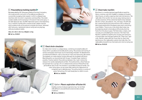
1 Pneumothorax training manikin 2 Chest tube manikin
Recommended by EMT (Emergency Medical Training) instructors, Now there is a manikin designed specifically to teach the
this model permits instruction of the delicate procedure for theory, anatomy, and skills needed to manage pre-hospital
successfully managing chest wounds in which a collapsed lung chest trauma, as well as ongoing chest tube maintenance. The
interferes with the victim‘s respiration and blood flow. The model right side of the manikin has two cut-away viewing areas to
accepts needle and thoracotomy in the second intercostal space, in provide awareness of the anatomical relationships between
the mid-clavicular line, the fifth intercostal space in the midaxillary the skin surface, musculature, ribs, and lungs. The left side
line. A realistic training aid to practice proper needle insertion has a pressurized tension pneumothorax site to relieve air
which enables built-up air pressure to escape the thorax. Delivered that accumulated within the pleural space and is restricting
with foot pump, replacement pleural cavity and carrying bag, lung inflation. There is also a site where chest tubes may
without procedural needle. be surgically placed to treat pleural effusion by draining
fluids from the pleural space. The fluid colour, volume, and
Size: 23 x 48.5 x 48.5 cm, Weight: 3.6 kg viscosity are controlled by the instructor. The Chest Tube
n Ref.no. R10037 Manikin is perfect for teaching the concepts and mechanics
of closed water-seal drainage systems like “Pleur-Evac” type
units. Manikin comes with two visual sites for chest tube
maintenance, five replaceable surgical chest tube sites, five
replaceable pneumothorax chest pads and a hard carrying case.
n Ref.no. R10130
3 Chest drain simulator
Our chest drain trainer is a unique design, combining simulated tactile soft
tissue within a practical and durable plastic casing. The practice and training of
clinical and surgical techniques can be realistically achieved. The identification
of anatomical landmarks, such as the sternal notch and intercostal (rib) spaces.
The students can make surgical incisions through the simulated soft tissue.
Blunt dissection can be practiced with forceps. Complete finger rotation is
possible. Practice realistic Pneumothorax between ribs 2 and 3 (where the
release of air can be clearly heard and felt). Pericardiocentisis, where fluid can
3 be aspirated into a syringe. This is to demonstrate removal of blood from the
pericardium. Ease of use: The simulator is accessible and easy to handle. The
outer casing opens via two metal clips making the replacement of
component parts relatively easy. The casing stands on a series of
sucker feet giving a firm and steady base on which to work. 2
n Ref.no. R10078
4 Option: Pleura aspiration effusion kit
Enables practice of pleural aspiration/tap. Can be filled
with different coloured fluids to demonstrate variable
conditions
4 n Ref.no. R10078-1
253