
Page 165 - EZ Catalog 18
P. 165
1
2
2 Baby Hippy
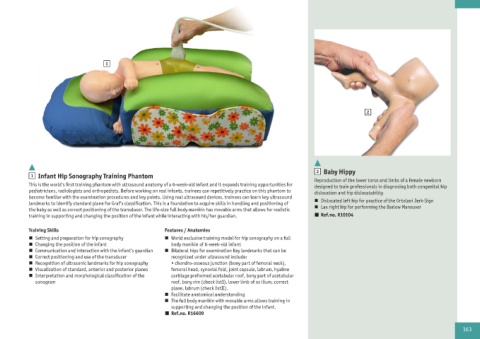
1 Infant Hip Sonography Training Phantom
Reproduction of the lower torso and limbs of a female newborn
This is the world‘s first training phantom with ultrasound anatomy of a 6-week-old infant and it expands training opportunities for designed to train professionals in diagnosing both congenital hip
pediatricians, radiologists and orthopedists. Before working on real infants, trainees can repetitively practice on this phantom to dislocation and hip dislocatability
become familiar with the examination procedures and key points. Using real ultrasound devices, trainees can learn key ultrasound
landmarks to identify standard plane for Graf‘s classification. This is a foundation to acquire skills in handling and positioning of n Dislocated left hip for practice of the Ortolani Jerk-Sign
the baby as well as correct positioning of the transducer. The life-size full body manikin has movable arms that allows for realistic n Lax right hip for performing the Barlow Maneuver
training in supporting and changing the position of the infant while interacting with his/her guardian. n Ref.no. R10104
Training Skills Features / Anatomies
n Setting and preparation for hip sonography n World exclusive training model for hip sonography on a full
n Changing the position of the infant body manikin of 6-week-old infant
n Communication and interaction with the infant‘s guardian n Bilateral hips for examination Key landmarks that can be
n Correct positioning and use of the transducer recognized under ultrasound include:
n Recognition of ultrasonic landmarks for hip sonography • chondro-osseous junction (bony part of femoral neck),
n Visualization of standard, anterior and posterior planes femoral head, synovial fold, joint capsule, labrum, hyaline
n Interpretation and morphological classification of the cartilage preformed acetabular roof, bony part of acetabular
sonogram roof, bony rim (check listI), lower limb of os ilium, correct
plane, labrum (check listII).
n Facilitate anatomical understanding
n The full body manikin with movable arms allows training in
supporting and changing the position of the infant.
n Ref.no. R16609
163