
Page 107 - EZ Catalog 18
P. 107
1 Gallstone Model
This half natural size model shows the anatomy of the
biliary system and ist surroundings in great detail. Both
the tissue changes caused by chronic inflammation and
acute inflammation (cholecystitis) are represented in the
gallbladder wall. Gallstones are shown in the following
typical places:
n spiral valve
n fundus of the gall bladder
n common bile duct
n papillary opening to the small intestine. 3
1
Mounted on base.
n Ref.no. K226
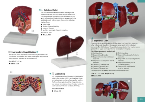
3 Segmental Liver
This reduced size model shows the division of the liver into 8 liver segments
after C. Couinaud. It explains the separate vessel supply of the individual
segments. The distribution of the portal vein separates the liver in a transverse
plane into an upper (cranial) and lower (caudal) Segment group.
2 Liver model with gallbladder The following segments are depicted:
2 n Left Liver Lobe n Right Liver Lobe
This realistic model reproduces a liver with the gall bladder. The n Segment I caudate lobe n Segment V caudal part of anterior
hilus vessels are shown as well as the extrahepatic ducts and the n Segment II cranial part of lateral segment
main ligaments. Mounted on removable stand. segment n Segment VI caudal part of posterior
Size: 16 x 12 x 11 cm n Segment III caudal part of lateral segment
segment
n Ref.no. K225 n Segment IV quadrate lobe n Segment VII cranial part of
posterior segment
n Segment IVa cranial part n Segment VIII cranial part of
n Segment IVb caudal part anterior segment
The model is not dissectible and comes on a removable stand.
4
4 Liver Lobule Size: 18 x 13 x 17 cm, Weight: 0.5 kg
This greatly enlarged model shows the fine detail of n Ref.no. K79
a single liver lobule, which is sectioned and shown
in relationship to portions of surrounding lobules. 3
The fine colouring distinguishes the portal veins
and vessels, venous sinusoids and central veins with
sections through the bile canaliculi. With key.
Size: 25 x 15 x 6 cm
n Ref.no. K78
105